Researchers at UTHealth Houston have received a five-year, $2.8 million grant from the National Institutes of Health to study the long-term effects of a fetoscopic spina bifida repair procedure. The funding will support follow-up for patients enrolled in the “Cryopreserved Human Umbilical Cord as a Meningeal Patch in Fetoscopic Spina Bifida Repair” trial until they reach 30 months of age.
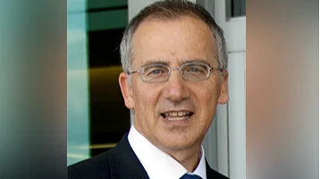
The study is led by Dr. Ramesha Papanna, professor at McGovern Medical School at UTHealth Houston and maternal-fetal surgeon at UTHealth Houston Fetal Center and Children’s Memorial Hermann Hospital. Researchers will examine neurological, motor, and developmental outcomes in relation to radiologic signs of spinal cord tethering among participants.
“It’s remarkable that an idea we had 13 years ago has become a therapy improving outcomes for children with spina bifida, helping them walk sooner,” said Papanna. “Advancing from bench concept to FDA phase 3 trial — backed by NIH grants — has been extraordinary. This new funding for the efficacy trial, already showing mobility gains, highlights the impact and promise for patients.”
Spina bifida affects about one in every 2,875 births annually in the United States. It is caused when the spine and spinal cord do not close properly during early pregnancy and can result in disabilities such as leg weakness or paralysis and bladder or bowel problems. The condition varies in severity: occulta (mild), meningocele (moderate), and myelomeningocele (severe). Treatment typically involves surgery along with ongoing medical care and physical therapy.
The UTHealth Houston team previously completed an early feasibility trial establishing safety for their approach and are now conducting late-stage testing. Their fetoscopic procedure differs from traditional open fetal surgery by using three small incisions rather than a large uterine opening; this allows most mothers to deliver vaginally unless otherwise indicated.
In this method, surgeons use a fetoscope—a high-resolution camera—and specialized tools to repair the defect through two layers: first placing a cryopreserved human umbilical cord patch over the spinal cord, then closing with skin tissue.
After birth, children return for follow-up assessments at 12 and 30 months old. These include neurological exams, developmental evaluations, and MRIs of both brain and spinal cord.
“Follow?up is essential, not only for each child’s care but also for the success of the study,” Papanna said. “These evaluations show us how children are progressing after this innovative therapy and help identify both positive outcomes and any areas that may need attention.”
The research team includes Stephen Fletcher (pediatric neurosurgery), Gabriel Anzueto (developmental pediatrics), Rajan Patel (neuroradiology), KuoJen Tsao (pediatric surgery), Jimmy Espinoza (OB-GYN), Sami Backley (maternal-fetal medicine), Peter Yang (pediatric neurosurgery), Jason Au (urology), Suzanne Lopez (neonatology), Lovepreet K. Mann (obstetrics/gynecology/reproductive sciences) and Dejian Lai (biostatistics).
Families or physicians interested in learning more about enrollment can visit the UTHealth Houston Fetal Center’s clinical trials page.